

- Ankle
- Bimalleolar fracture
- Fibular fracture
- Osteotomy
- Subcutaneous emphysema
- Tibial pilon fracture
- Trimalleolar fracture
- Unimalleolar fracture (lateral)
- Arm
- Humeral shaft fracture
- Nightstick fracture
- Proximal humerus fracture
- Pt 8: Investigation
- Pt 9: Investigation
- Pt 10: Preoperative evaluation
- Pt 10: Postoperative control
- Pt 16: Investigation
- Torus of the distal radius
- Elbow
- Capitellum fracture
- Condyle fracture
- Distal humerus fracture
- Elbow dislocation
- Intercondylar fracture
- Intra-articular swelling
- Monteggia fracture
- Olecranon fracture
- Radial head fracture
- Supracondylar fracture
- Trochlear fracture
- Foot
- Calcaneus fracture
- Clubfoot
- Cuboid fracture
- Enthesopathy
- Hallux valgus
- Jones fracture
- Lisfranc fracture
- Metatarsal fracture (1st)
- Metatarsal fracture (4th)
- Metatarsal fracture (5th)
- Metatarsophalangeal luxation
- Metatarsus varus
- Osteomyelitis
- Phalangeal fracture
- Pseudo-Jones fracture
- Rheumatoid arthritis
- Sesamoid luxation
- Talus fracture
- Hand
- Bennett fracture
- Boxer's fracture
- Interphalangeal luxation
- Metacarpal fracture (4th)
- Metacarpal fracture (5th)
- Metacarpophalangeal luxation
- Phalangeal arthrosis
- Radiocarpal arthritis
- Reverse Bennett fracture
- Rheumatoid arthritis
- Rhizarthrosis
- Rolando Fracture
- Hip
- Acetabular fracture
- Pt 11: Preoperative evaluation
- Pt 11: Postoperative control
- Pt 13: Investigation
- Pt 65: Investigation
- Avascular necrosis
- Coxa magna
- Coxa plana
- Coxarthrosis
- Pt 19: Preoperative evaluation
- Pt 72: Investigation
- Pt 78: Preoperative evaluation
- Pt 78: Postoperative control
- Pt 79: Preoperative evaluation
- Pt 79: Postoperative control
- Pt 80: Preoperative evaluation
- Pt 80: Postoperative control
- Developmental dysplasia of the hip
- Femoral head fracture
- Femoral head fracture-dislocation
- Femoral neck fracture
- Pt 17: Preoperative evaluation
- Pt 17: Postoperative control
- Pt 18: Preoperative evaluation
- Pt 18: Postoperative control
- Pt 18: Postoperative control
- Pt 19: Preoperative evaluation
- Pt 19: Postoperative control
- Pt 20: Preoperative evaluation
- Pt 20: Postoperative control
- Pt 21: Preoperative evaluation
- Pt 21: Postoperative control
- Heterotopic calcification
- Intertrochanteric fracture
- Pt 13: Investigation
- Pt 22: Preoperative evaluation
- Pt 22: Postoperative control
- Pt 23: Preoperative evaluation
- Pt 23: Postoperative control
- Pt 24: Preoperative evaluation
- Pt 24: Postoperative control
- Legg-Calvé-Perthes disease
- Os acetabuli
- Periprosthetic fracture
- Slipped capital femoral epiphysis (SCFE)
- Subtrochanteric fracture
- Total hip prothesis dislocation
- Knee
- Bone graft
- Enchondroma
- Femoropatellar arthrosis
- Gonarthrosis
- Pt 82: Investigation
- Pt 82: Postoperative control
- Pt 83: Preoperative evaluation
- Pt 83: Postoperative control
- Pt 84: Preoperative evaluation
- Pt 84: Postoperative control
- Intra-articular swelling
- Pt 44: Preoperative evaluation
- Pt 70: Preoperative evaluation
- Pt 82: Investigation
- Pt 83: Preoperative evaluation
- Kirschner wire
- Osteochondral lesion
- Osteochondritis dissecans
- Osteophytes
- Patellar fracture
- Tibial plateau fracture
- Pt 35: Preoperative evaluation
- Pt 35: Postoperative control
- Pt 36: Preoperative evaluation
- Pt 36: Postoperative control
- Pt 50: Preoperative evaluation
- Pt 50: Postoperative control
- Tibiofemoral luxation
- Leg
- Femoral shaft fracture
- Pt 42: Preoperative evaluation
- Pt 42: Postoperative control
- Pt 43: Preoperative evaluation
- Pt 43: Postoperative control
- Fibular fracture
- Fibular shaft fracture
- Maisonneuve fracture
- Phlebolith
- Tibial shaft fracture
- Neck
- Edema
- Halo
- Pelvis
- Ischial enthesopathy
- Ischiatic ramus fracture
- Pubic ramus fracture
- Sacral fracture
- Vascular calcifications
- Shoulder
- Acromioclavicular arthrosis
- Acromioclavicular luxation
- Pt 88: Investigation
- Pt 91: Preoperative evaluation
- Pt 91: Postoperative control
- Pt 92: Investigation
- Clavicle fracture
- Glenohumeral arthrosis
- Glenohumeral dislocation
- Hill-Sachs fracture
- Rotator cuff tear
- Scapular fracture
- Spine
- Burst vertebral fracture
- C1 fracture
- C1-C2 instability
- C5 fracture
- Cervical fracture-dislocation
- Discarthrosis
- Facet arthrosis
- Flexion-distraction fracture
- Forestier disease
- Odontoid fracture
- Retrolisthesis
- Rheumatoid arthritis
- Scoliosis
- Spondilolysthesis
- Uncarthrosis
- Wedge fracture
- Thorax
- Rib fracture
- Wrist
- Avulsion fracture of the ulnar styloid
- Pt 11: Postoperative control
- Pt 31: Preoperative evaluation
- Pt 57: Preoperative evaluation
- Pt 57: Postoperative control
- Barton's fracture
- Chauffeur's fracture
- Colles fracture
- Pt 30: Preoperative evaluation
- Pt 30: Postoperative control
- Pt 31: Preoperative evaluation
- Pt 31: Postoperative control
- Distal radius fracture
- Galeazzi fracture
- Perilunate fracture-dislocation
- Physeal injury of the distal radius
- Radial styloid fracture
- Radioulnar joint disruption
- Reverse Barton's fracture
- Scaphoid fracture
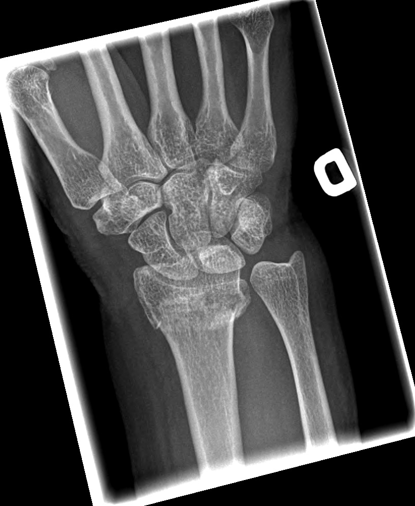
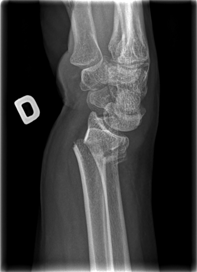
62-year-old female:
Right wrist pain and deformity following a fall from standing height on a dorsiflexed wrist.
Right wrist pain and deformity following a fall from standing height on a dorsiflexed wrist.
Colles fracture
Bony irregularities
Slightly comminuted distal radius fracture with dorsal tilt.
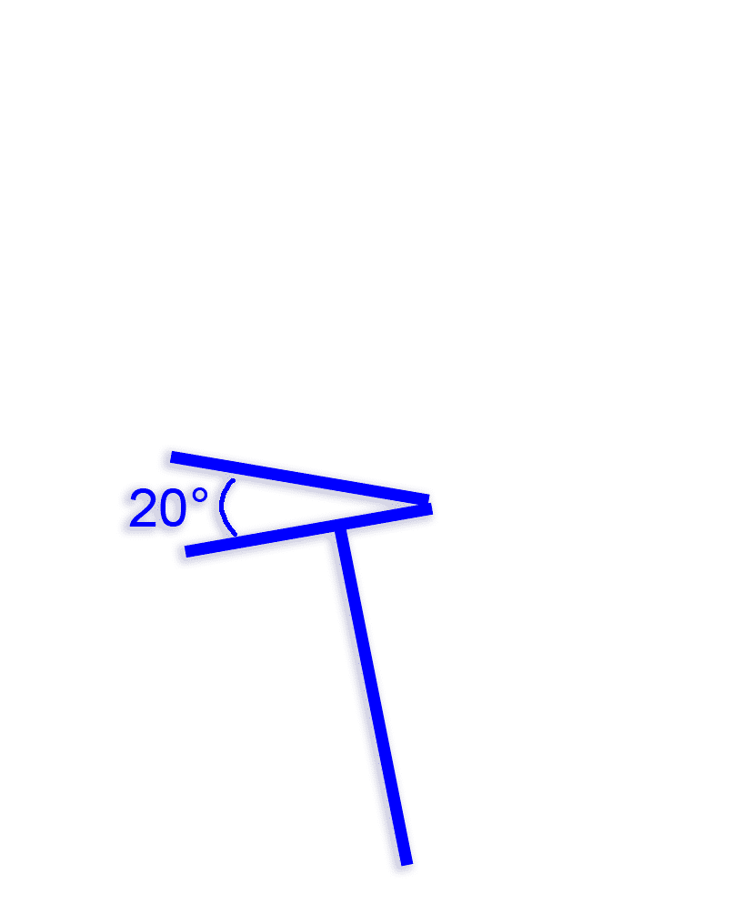
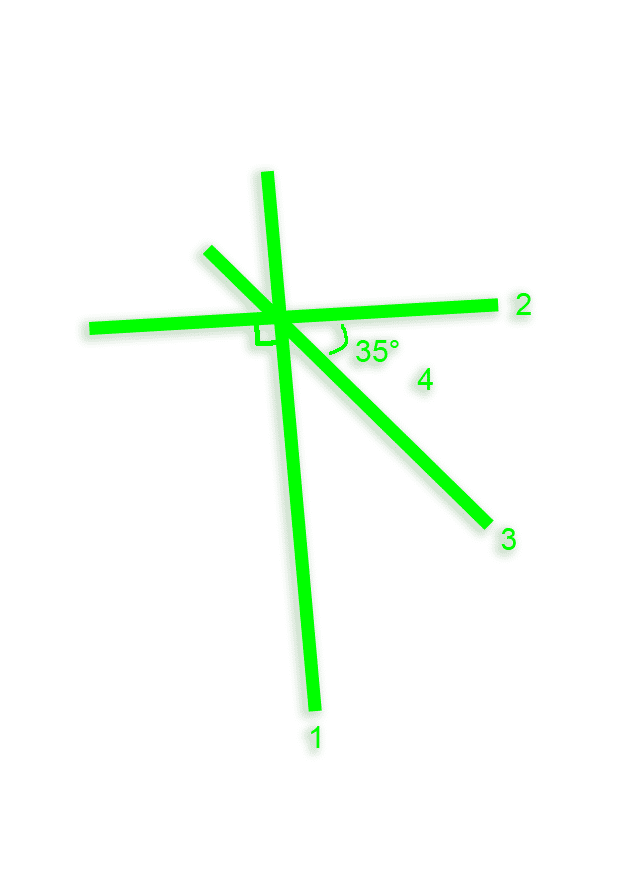
Volar tilt
You can follow the steps from 1 to 4 to measure the volar tilt. Normally, the distal radius (line 3) is angulated 10-15° volarly on the lateral view compared to line 2, which is orthogonal to line 1. Here, this angulation is reversed dorsaly 35° (step 4: angle between line 2 and line 3).
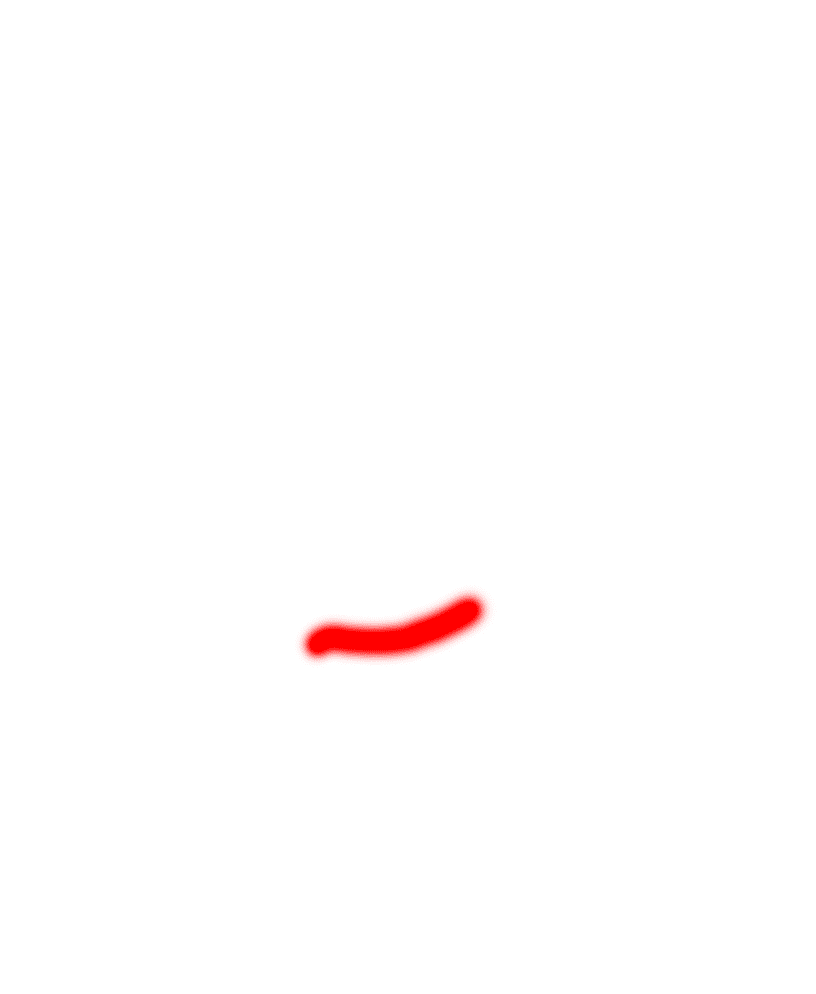
Radial tilt
Angulation measurement
The radial tilt is about 20°.
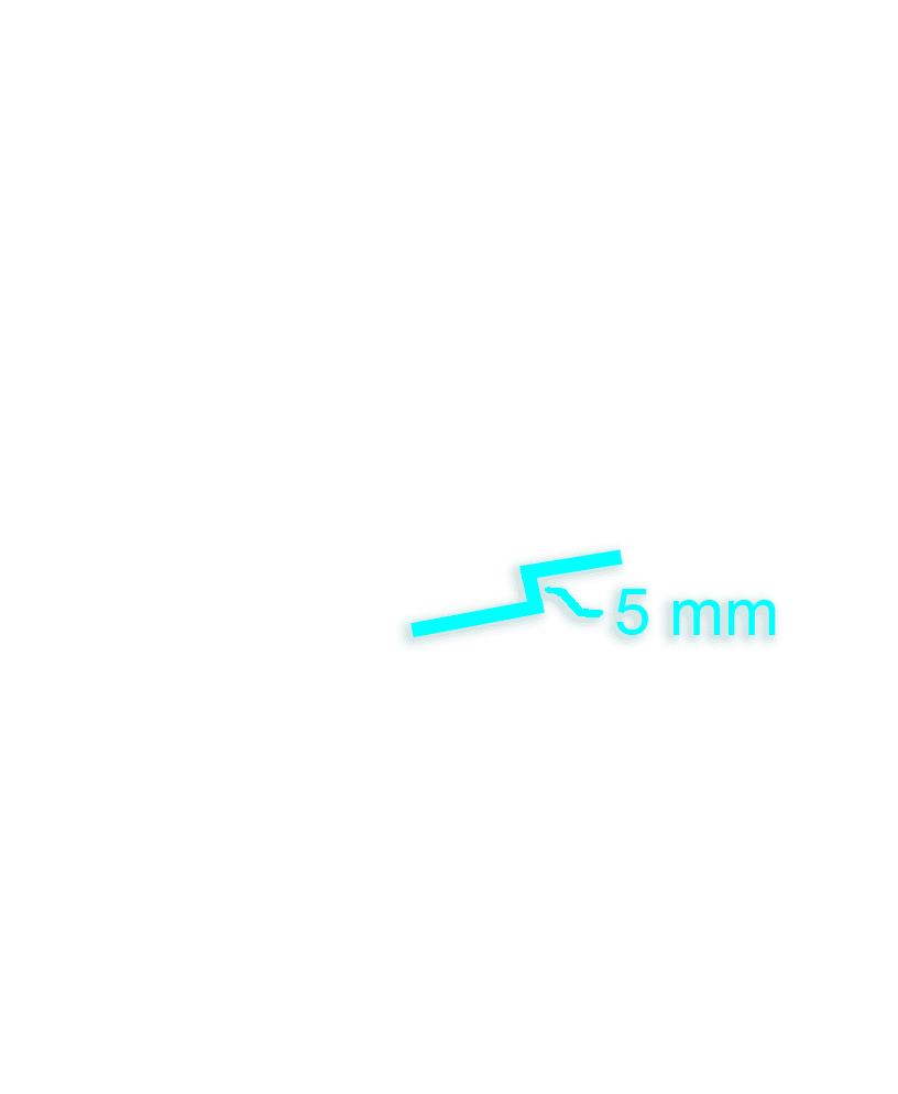
Ulnar variance
The ulnar variance is positive at 5 mm (there is a shortening of the radius). The ulnar variance is considered positive when the ulna is longer than the radius.
Intact articular surface
The articular surface is smooth: the fracture appears extraarticular on these radiographs.
Related chapters
- Atelectasie
- Pneumonie

Zoom:

test


Zoom:
test
.jpg
.png
154
2
5
../ortho/resources/studies/
../sandbox/